April 26, 2019, by Kathryn Steenson
MRI Collections Project: Sir Peter Mansfield’s research papers
A significant proportion of Sir Peter Mansfield’s papers, as held by Manuscripts and Special Collections, relate to his own studies and research. Like many academics, Sir Peter kept his A’Level and undergraduate notebooks, and these provide illuminating insights into Physics education in the 1950s. Sir Peter did his undergraduate degree in Physics at Queen Mary College, London, from 1956 to 1959.
Sir Peter’s research notes and papers within the collection date from 1959, when he started studying for his PhD on ‘Proton Magnetic Resonance Relaxation in Solids by Transient Methods‘ (also at Queen Mary College), up to around 2000. They include notebooks and files relating to his early experiments on nuclear magnetic resonance, often with photographs of experimental results (in the form of electronic signal traces) glued into them. As his research progressed, such photographs gradually gave way to images of MRI scans as we would recognise them today. The collection also bears witness to the variety of Sir Peter’s research interests, ranging from establishing the basic principles of MRI in the 1970s, through his work on Echo Planar Imaging and real time imaging in the 1980s, to acoustic screening (i.e. reducing the noise of the MRI process) and fluid transport in porous rocks in the 1990s. Also present in the collection are files in which he gathered together academic papers on a particular topic, then added his own notes. Topics he studied in this way include Fast Fourier Transforms, Liquid Crystals, Cross Relaxation, Chemical Shift and Neural Stimulation. The collection also includes numerous handwritten and typescript drafts of many of his published academic papers, including diagrams and photographs.
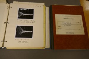
Examples of a ringbinder and a notebook from the 1960s in which Sir Peter recorded his experimental work.
Sir Peter documented his work in various ways. Many of his early mathematical proofs relating to nuclear magnetic resonance are written on loose sheets of unlined paper, whereas his experimental work is generally recorded in bound notebooks or ringbinders. In the 1970s he even used two large diaries to write sets of notes and calculations, instead of the more usual appointments. He also sometimes stored loose sheets of notes between the pages of his notebooks and files, which makes life more challenging for the archivist!
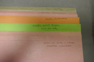
Examples of some of the files in which Sir Peter stored research notes on particular topics.
Some of these files can contain a wide range of materials such as handwritten notes and diagrams on paper, photographs of experimental results, published academic papers, and overhead projector transparencies which would have been used to deliver talks or lectures.
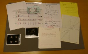
An example of the variety of materials found in one of Sir Peter’s research files.
The collection also contains evidence of how Sir Peter and his group used computers in the 1970s and 1980s. Some files include printouts of computer programs and their results, on topics such as calculating the magnetic fields generated by different types of coil, or the programming of equipment to generate a specific sequence of electronic pulses. These programs date from the time when even computers with tiny amounts of memory (by today’s standards) were expensive. For example, in his autobiography, Sir Peter refers to how he used some grant money in 1971 to purchase a Honeywell computer with just 4Kb of magnetic memory! This collection therefore acts as a salutary reminder of how scientific research was done before desktop PCs and electronic storage became cheap and ubiquitous.
We are currently in the process of cataloguing all of these research notes and papers. Once completed, they will form a fascinating resource for those interested in the scientific process and the development of medical technology.
No comments yet, fill out a comment to be the first


Leave a Reply