March 22, 2019, by Brigitte Nerlich
Talking organelles: A riot of metaphors
A few days ago, somebody tweeted an article on organelles and somebody else tweeted an article on how worms regenerate their bodies. I was just in a slump of Brexit malaise when I saw this and thought, “oh, there is life outside Brexit at least in worms and cells”. So, I started to read an article published in Nature by Elie Dolgin, a science journalist: “How secret conversations inside cells are transforming biology”. What’s discussed in this article is probably well-known to cell biologists, but it wasn’t to me.
I was hooked, not only because of the science but also, and perhaps even more so, by the firework of metaphors that I encountered. This made me think again about the fact that science/communication without metaphor is basically impossible. This seems to be particularly the case for cell biology, as the recent book by Andrew Reynolds has shown, entitled: The Third Lens: Metaphor and the Creation of Modern Cell Biology.
In the following I shall put the Nature article under metaphorical microscope and tease out various types of metaphors used in it: conventional metaphors, more creative metaphors, and finally an extended metaphor, telling a whole new story about organelles.
I shall also say something about the interactions, indeed, ‘cross-talk’, between verbal and visual images, which was important for advancing research into organelles, leading up to discoveries relating to how organelles themselves talk between each other.
What are organelles?
The Oxford English Dictionary defines organelles as: “Any of various discrete, usually membrane-bound, structures within a cell or unicellular organism that perform specialized functions”.
The term is first attested for 1915 but harks back to an earlier term organella/organula from 1889. Somebody writing in American Naturalist pointed out: “It may possibly be of advantage to use the word organula here instead of organ, following a suggestion by Möbius. Functionally differentiated multicellular aggregates in multicellular forms or metazoa are in this sense organs, while for functionally differentiated portions of unicellular organisms or for such differentiated portions of the unicellular germ-elements of metazoa the diminutive—organula—is appropriate.”
Ten years later, in 1896, as Dolgin points out in her Nature article, “cytologist Edmund Beecher Wilson drew the cell with organelles neatly tucked into their own distinct cytoplasmic compartments” (p. 164). Here it is: Image from his textbook “The cell in Development and Inheritance”, second edition, 1900 (Wikimedia Commons):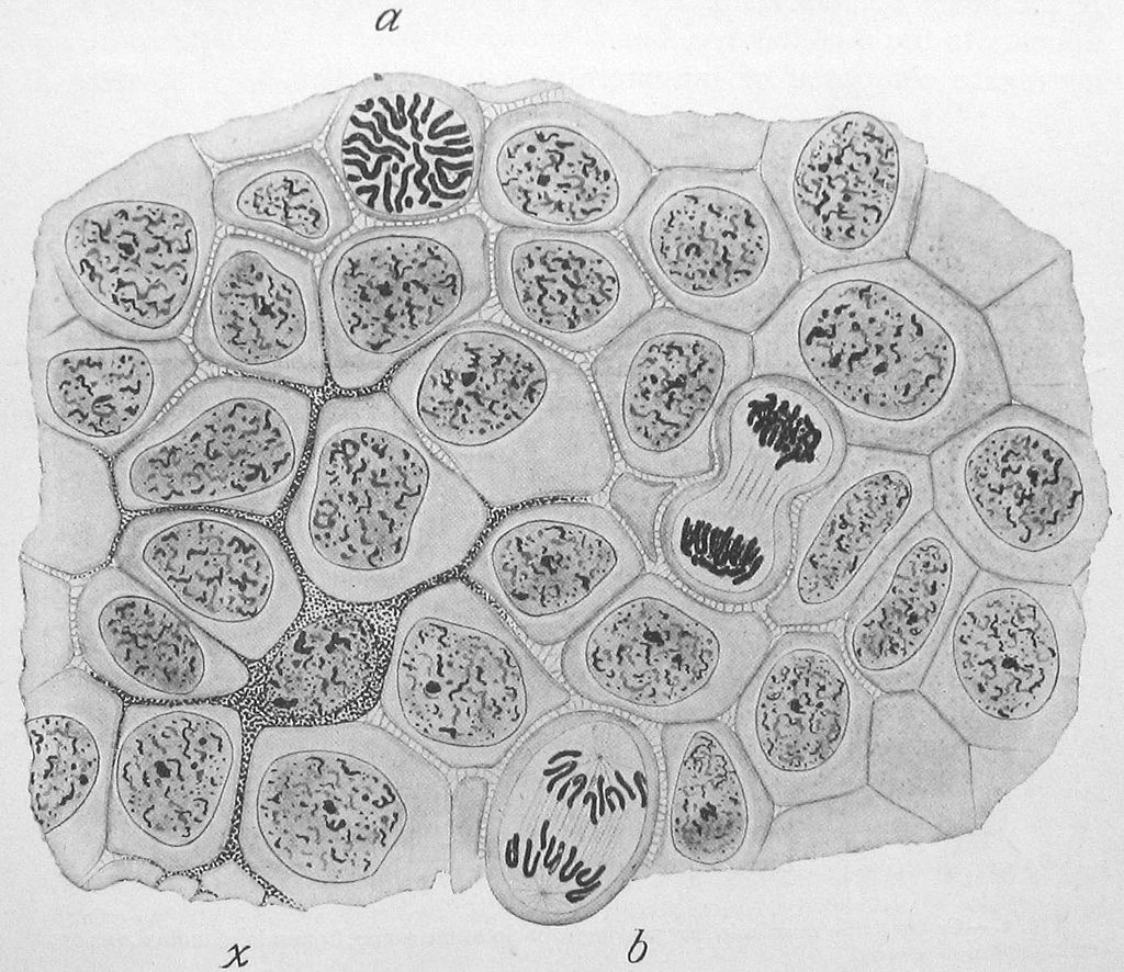
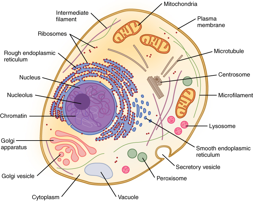
In biology textbooks the various organelles inside a cell are generally depicted and labelled in standard ways, as for example in the featured image for this blog post. One can also often find tables, like this, that list organelles and their functions. And in the description of functions, metaphors creep in – in this case we are dealing with a eukaryotic cell. The metaphors in this table are quite tame: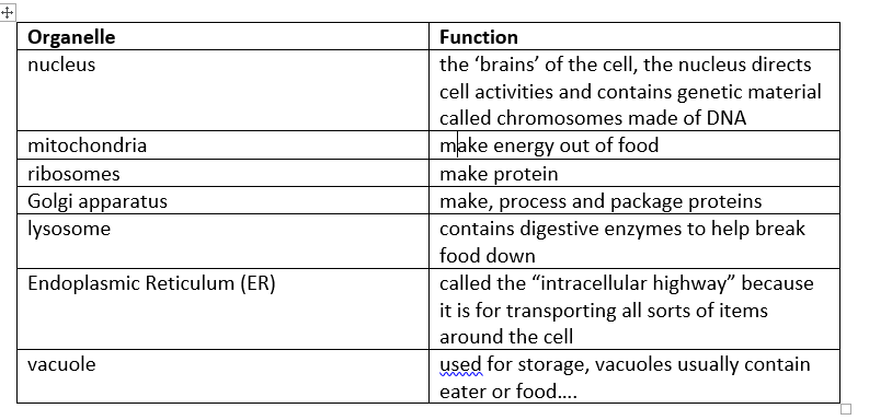 In other textbooks we find other metaphors and they circulate in science talk and text. When I tweeted about my intention to write this post, Matthew Cobb tweeted: “”powerhouse” (mt), “construction yard” (ER), “warehouse” (Golgi), “vacuum cleaners” (lysosomes), “dustbin” (vacuole), “frontier” (cell wall)”.
Some of these metaphors are more conventional than others, I suppose. The most overused and clichéd one seems to be that of ‘powerhouse’ for mitochondrion. As Liam Drew pointed out in a tweet: “I wrote something like ‘mitochondria – the powerhouses of the cell as science writers are contractually obliged to describe them’ and my copy editor changed it to ‘mitochondria – the powerhouses of the cell’”.
Back to the article under my microscope. The Nature article uses the conventional metaphor of ‘science as a journey’ through which we are told the story of Jean Vance who, after hitting a series or ‘roadblocks’ over a long period of time, finally published a ‘landmark’ paper which put organelle ‘conversations’ ‘on the map’ (that last one is my own metaphor).
I won’t go into this rather fascinating story, but focus instead on the metaphors for organelles and what they do that we encounter on the way. I just want to stress however that this article tells a very typical story about how science is often ‘done’ (it all starts with “I thought I’d made a big mistake” p. 162). It is also a typical example of how science is communicated: using well-established metaphors, extracting metaphors from scientists who use the ones they always use or create new ones on the spot, and giving that mix some creative twirls.
Conventional metaphors
As we have seen, there are lots of more or less conventionalised metaphors floating about that can be used to talk about organelles. Some are indispensable to science, what one can call theory-constitutive metaphors, some are didactic, some are explanatory, some are so clichéd that one has to cringe etc. The article under the microscope doesn’t use all of them, only some. Here they are: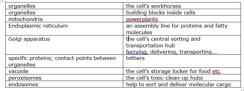
Creative metaphors and similes
These are not the only metaphors used by/collected by Elie Dolgin. She also uses more creative ones, as in the title (“The secret conversations inside cells: Organelles – the cell’s workhorses – mingle much more than scientists ever appreciated”) and also in the section headings, which also use alliteration: “first contact”, “tether together”, “function junction”, “bad dancers” – we’ll come back to the dance metaphor! Here is a list of the creative metaphors and comparisons (similes) I found: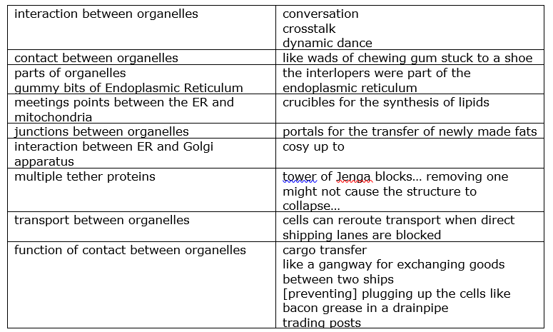
Extended metaphor
Now we come back to the dance metaphor, because one scientist interviewed for this article uses it as an extended metaphor to tell a whole new story about organelles.
“Gökhan Hotamışlıgil, a metabolic-disease researcher at the Harvard T. H. Chan School of Public Health in Boston, Massachusetts, likens the relationship between the ER and mitochondria to a sensual and dynamic flamenco performance. Just like dancers, the organelles ‘contact and separate, and then come into contact again, and flirt a little bit and go away’, he says. But in diseased liver cells, the two organelles stay entwined, and the rhythm is sluggish. ‘It doesn’t look very elegant,’ says Hotamışlıgil, who has shown that excessive contact between the ER and mitochondria in mouse liver cells is linked to insulin resistance, diabetes and obesity. ‘You can’t slow-dance flamenco — and that’s how the mitochondria–ER relationship becomes under metabolic stress,’ he adds.”
Verbal and visual metaphors
I have now dissected this great article in terms of the metaphors it uses. While doing so, it became clear that focusing on the verbal is not enough and that images and visual metaphors also play an important role in doing organelle science and communicating organelle science, from the end of the 19th century onward. The article itself contains a wonderful little cartoon of dancing organelles but also a (I believe) video still of the Endoplasmic Reticulum ‘swapping goods with various organelles’.
In fact, video footage of organelles doing their stuff was important in persuading sceptics that organelles are in constant conversation with each other. Images or pictures also made scientists themselves enter into conversation with each other: “scientists studying vesicles did not generally communicate with those who specialize in signalling through calcium ions. ‘There was no contact in the contact field,’ Levine says.” (p. 163)
Images changed from drawings to traditional microscope images to “super-resolution light microscopy to capture kaleidoscopic-colour 3D movies of intricate interactions between six organelles: the ER, mitochondria, the Golgi complex, peroxisomes, lysosomes and fat deposits called lipid droplets” (p. 164). Some researchers made “black-and-white films that showed the ER ensnaring mitochondria and other organelles in mouse neurons” (p. 164). Here we see a nice coming-together of verbal and visual metaphors.
However, as Dolgin points out, text book images of organelles have changed little since 1896. “From the ER to the Golgi to the vacuole to the endosome, each organelle is still shown in isolation, not as a dynamic dance of parts that continuously embrace and separate. ‘Nothing is drawn the way the cell actually looks,’ says Voeltz. ‘It would be nice to update that image.'”
Do we also need to update the language? It seems to work quite well at the moment, I think.
Featured image: Animal cell and components (Wikimedia Commons)
No comments yet, fill out a comment to be the first


Leave a Reply