March 13, 2019, by Dr. Meghan Gray
CSI: East Midlands
In this guest post by Dr. James Sharp, we learn about about cutting-edge new techniques used to measure fingerprints. A paper describing this work was published in the journal Science and Justice and was recently featured on BBC CrimeWatch Roadshow. For more information on how physics and forensics go together, see a previous post on this blog featuring Dr. Sharp’s work on footprint detection.
Retrieval of fingerprint evidence from metal surfaces can be tricky. Under ideal conditions, the development of what are called latent (or hidden) fingerprints involves application of some kind of chemical agent (e.g. superglue fumes and/or fluorescent dyes) that is designed to make the fingerprint easier to visualise. Photographs of the freshly developed prints can then be taken and compared to the fingerprints of individuals who may be linked to a crime scene. However, the optical properties of metals prevent clear visualisation of latent prints even when these contrast-enhancing techniques are used.
Things are further complicated when the item being used to recover evidence is something like a knife blade or a bullet casing as these can often be exposed to harsh environments that result in damage to, or erosion of the fingerprints. In the case of knife blades, these are often left lying around at a crime scene and can be exposed to the elements. Quite quickly, rain and mechanical contact can have the effect of damaging any prints that might be on the item. Similarly, bullet casings are exposed to extremely high temperatures, pressures and mechanical friction during firing and this often obliterates the majority of any fingerprints that may have been left on them. In many cases, only residual traces of fingerprints may be present on these items and conventional development techniques struggle to produce any useful images.
In a recent collaboration with the University of Derby, a UoN Physics undergraduate summer student (Adam Reeve) and PhD student (Tshaiya Thandauthapani) performed a comparative study of conventional fingerprint enhancement techniques with a highly sensitive and non-destructive surface science technique called Time of Flight Secondary Ion Mass Spectroscopy (ToF SIMS). The ToF SIMS instrument that was used for this study is currently based in the School of Pharmacy at the University of Nottingham.
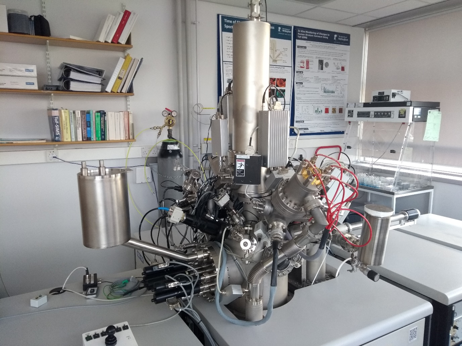
The ToF SIMS Instrument based in the School of Pharmacy at the University of Nottingham that was used to image fingerprints on metal surfaces.
ToF SIMS uses an ion beam to scan a surface such as metal substrate, a knife blade, or even a bullet casing. This primary ion beam causes the ablation of ultrathin layers of material (typically only a molecule or so thick) and the charged molecular fragments that are emitted from the surface are analysed and sorted by their mass-to-charge ratio. The molecular fragments that are emitted are individual to and characteristic of different materials. Scanning the primary ion beam across the surface and looking at the molecular fragments that are emitted therefore allows an image or map of the different chemical species that may be present on a surface to be built up. This technique was used in our recent study to look for the oily residues and salts that are secreted in fingerprint sweat and to form images of fingerprints on these problematic surfaces. The image below shows an example of a fingerprint that was extracted from a stainless steel surface (one of the most difficult substrates) using the ToF SIMS technique along with a comparison of optical images taken after using conventional enhancement techniques. The ToF SIMS images show clear evidence of fingerprint ridge structure and even detailed information about the location and shape of sweat pores, where the conventional images show no evidence at all of a fingerprint being present.
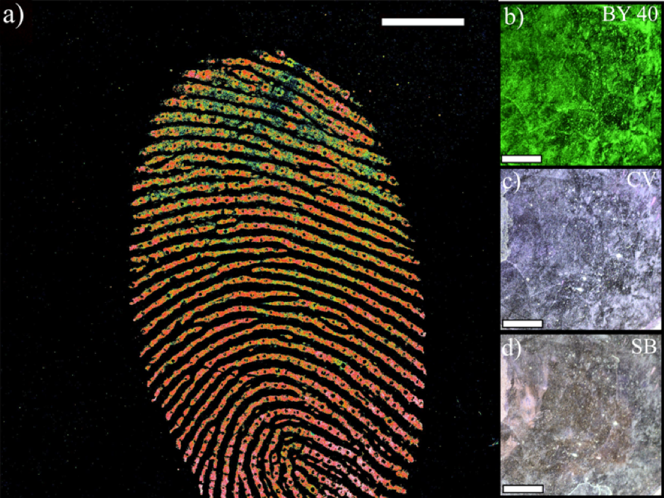
Fingerprints on stainless steel. Panel a) A ToF SIMS image of the surface of a stainless steel sample. This a combined image of the intensities/concentrations of sodium and potassium ions on the surface. Panels b-d) show optical images taken from the same region of the substrate containing the fingerprint after superglue fuming and exposure to fluorescent dyes Basic Yellow 40 (BY40), Crystal Violet (CV) and Sudan Black (SB).
No comments yet, fill out a comment to be the first

Leave a Reply