March 2, 2018, by jicke
Plant cell research gets Gold!
Innovative plant research from a team of scientists from the University of Nottingham has been given a gold prize for best original article by The Plant Journal.
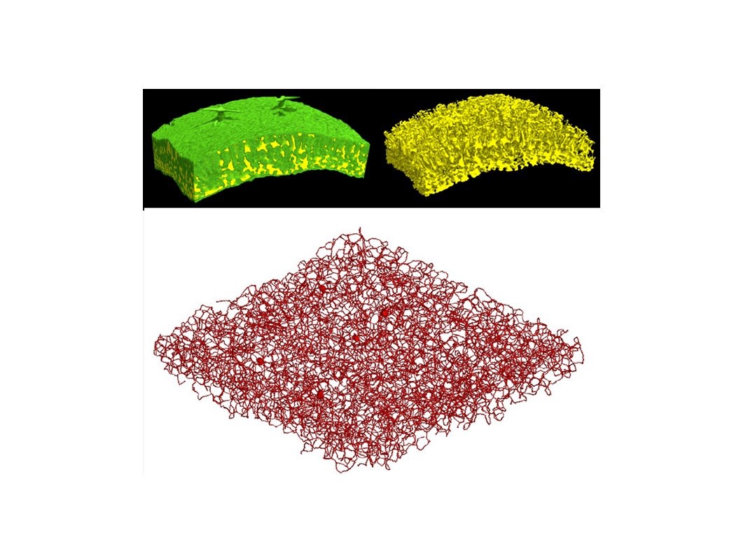
An editorial board at The Plant Journal voted for the winning research article – Cell density and airspace patterning in the leaf can be manipulated to increase leaf photosynthetic capacity. The research, published in September, was a collaboration between soil scientists in Nottingham and plant scientists in Sheffield. The team used innovative imaging techniques to explore the cell density and airspace patterning in plants and how this can be manipulated to increase photosynthesis.
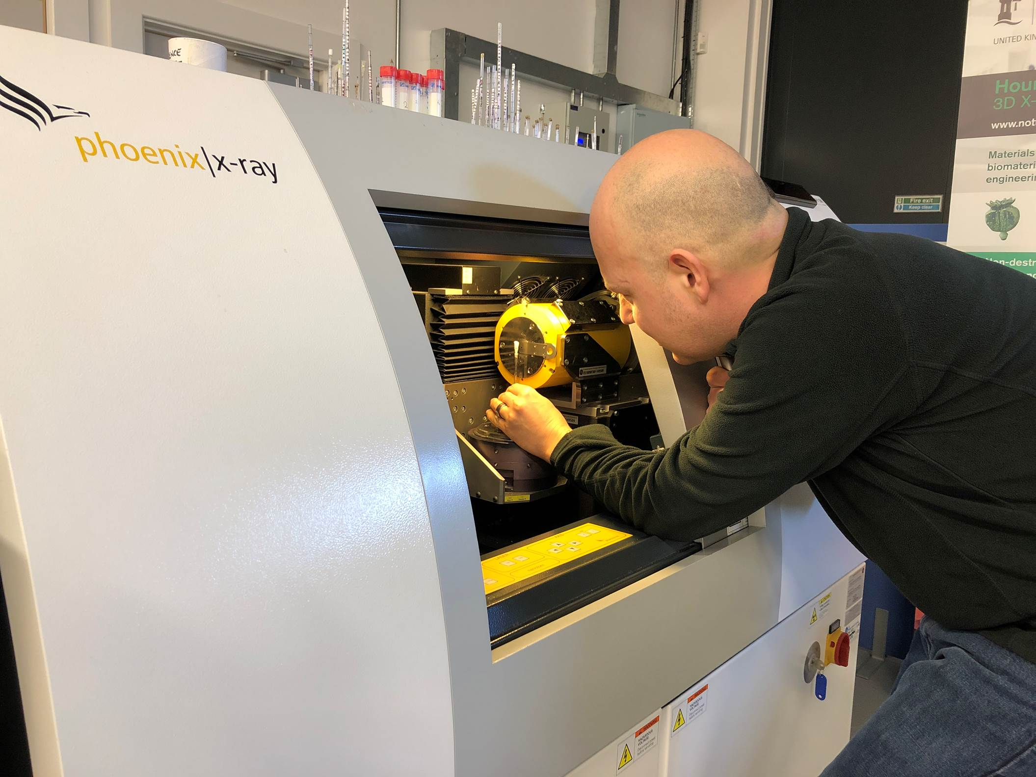
Professor Sacha Mooney worked with Dr Craig Sturrock and Dr Andy Mathers on the research using CT Scanning techniques to carefully examine leaf cell structure. Professor Mooney says: “We’re delighted to have been given this award by the Plant Journal. This was an exciting piece of work, particularly as we were able to translate our expertise in soil research and the analysis of plants – the analysis we did on the cell network in leaves is very similar to that which we carry out on the pore network in soils.”
Improving photosynthesis
Through meticulous imaging and analysis of the cell structure and the pattern of cell division, growth and separation during leaf development, the team found that cell density can be increased to engineer improved photosynthesis, which can boost the growth and health of plants.
Professor Mooney continues: “Understanding how the plant cells work to produce photosynthesis means we can now see that it is a process that can be manipulated, this could allow us to develop better growing techniques and capabilities which could potentially have a huge impact on improving crop yields and plant performance.”
Hounsfield Facility
The research was carried out at the Hounsfield Facility that is equipped with three CT scanners — capable of imaging objects as fine as a soil particle or a root hair to a fully mature root system. With these scanners and specialised image analysis methods Nottingham researchers can image the structure of plant roots in a non-destructive way growing through soil throughout the life of the plant — from seed to flowering.
Imaging the hidden half of plants
There’s a small high resolution scanner for visualising fine details such as single roots, root hairs and the soil around them: a medium scale micro CT scanner to image an entire root system: and a large custom designed CT system to look at plants such as wheat throughout its whole growth cycle from seed to flowering — bringing the field closer to the lab than ever before.
X-ray CT produces a 3D image of the scanned sample. The RooTrak image analysis software, developed by experts in the University’s School of Computer Science, identifies root material within those images and builds a 3D model of the root system that can be viewed from any angle.
No comments yet, fill out a comment to be the first


Leave a Reply