July 4, 2010, by Teaching at Nottingham
Development of Anatomy CAL using Xerte Online Toolkits
The Medical School had a number of Anatomy CAL (Computer Aided Learning) packages, created in Authorware over a number of years. Due to their age and format they were not easy to update or support but they did contain relevant and useful content. There was a requirement for the packages to be reviewed and where appropriate new versions created in a format that would be easy to update and easy for students to access. A Teaching and Learning funded project ran for 6 months in the first half of 2010 to address this.
Implementation
The old CAL packages were reviewed and a list of new packages that were required was created. The list included areas of anatomy such as Cranial Nerves and Joints of the lower limb. Xerte Online Toolkits was used to create the new interactive learning material that could be accessed by students over the web. The new CAL can be viewed at any time and any place by students with a PC with an internet connection. Some of the features in Toolkits were modified or repurposed to better meet the Medical School Anatomy requirements. An example of this was the ability to specify the shape of a hotspot so that it could follow the irregular shapes found in human anatomy. This was essential for the use of the interactive components available in Toolkits.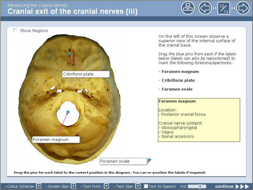 Fig 1.
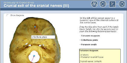
The CAL packages contain core content, interactive activities and self-assessment questions. An example of an interactive activity included in the anatomy CAL is ‘drag and drop labelling’. Students are asked to correctly label parts of an image by dragging labels to the correct position. The example in Fig.1 shows an image of the cranial base and some of the areas that need to be selected are quite small and would be difficult to locate with a large label. Toolkits has a pin for each label which make it suitable for use with small target areas. The student has to drag a pin rather than the whole label to a precise area of the graphic. The pins only lock into place when they are dragged to the correct position and details about the selected part of the cranial base are displayed in a yellow box as shown in the graphic in Fig.1.
Another interactive feature is seen in the ‘categories’ activity which asks student to drag labels into the correct category. Fig. 2 shows an example of this where students are asked to drag labels for different cranial openings to the correct entry/exit points for cranial nerves V and VII. The labels have been dragged by a student who has then clicked on the ‘Check Answers’ button. The correctly placed labels are shown with a tick and students are able to move incorrectly placed labels and check their answers again.
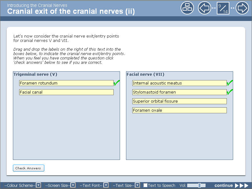 Fig 2.
Medical students can access the new CAL through the NLE (Networked Learning Environment) as part of the relevant module.
Student feedback
A student evaluation took place as part of a pilot. They commented that they liked being able to do things online that were close to what they are assessed on. They particularly liked the labelling and categorisation activities (as shown in the above examples). They felt that the interactive activities were a real strength of the online learning and had benefits over paper based materials.Reflections on the project
Storyboarding was a very useful step in the project as this created a picture of how the completed learning material would look. Modifications could then be made before spending time developing the material. As Toolkits supports a number of interactive activities, getting someone to explain how they work can save a lot of time. The end product of these activities was better than expected.The work on this project has led to work starting on the creation of short tutorials for students to complete in preparation for attending the dissection room sessions.
Dr Deborah Merrick and Terrence Mayhew
School of Life Sciences
No comments yet, fill out a comment to be the first


Leave a Reply